Twins and infertility
Keywords: equine, infertility, twins, ovulation, abortion.Note: the term conceptus is used here by the author, in preference to embryo. In the author's mind, conceptus refers to the embryo together with its membranes while embryo refers to the future fetus. Also, because of the importance of twinning in equine reproduction, the author has included more text than usual in this entry.
Twin preovulatory follicles seen on ultrasound.

Image size: 900 x 694px
Conventional wisdom once dictated that mares should not be bred when twin preovulatory follicles were present. This was based on the fact that twins are a common cause of abortion in mares. With the increase in knowledge that came with common use of ultrasonography, this philosophy has changed. Today, it would be considered poor management to miss this breeding opportunity. Indeed, if mares where not to be bred whenever twin follicles were present, perhaps 15 to 20% of all breeding opportunities would be lost. This is especially the case in breeds such Thoroughbreds and Standardbreds where twin ovulations are common. It is also significant in any breed with the approach of the summer solstice because day length and ovulation rate are positively correlated.
Current philosophy dictates that mares should always be bred when twin follicles are present. Indeed, even when twin pregnancies are not discovered, the overall singlet pregnancy rate is higher when this approach is used.
Notes on the timing of pregnancy diagnosis:
When two conceptions occur during a single estrous period, one conceptus will invariably be older and larger than the other conceptus when pregnancy diagnosis is performed. This is important to bear in mind. Although not common, twin ovulations can be separated by several days, making it possible to miss a younger conceptus when routine pregnancy diagnosis is performed at 14 to 15 days after ovulation.
Newer, high resolution ultrasound units make it possible to see pregnancies at 12 days or even earlier. If there a second, younger conceptus in the uterus at this time, it may not be seen. However, if pregnancy diagnosis is delayed to 14 days, it is very likely that both conceptuses will be seen. After 16 days, embryo movement within the uterus will have ceased and the opportunity to crush one embryo without harming the other will have diminished significantly. Therefore pregnancy diagnosis should be scheduled at 14 to 16 days after the time that the first of two intact (preovulatory) follicles was last seen. This is explained below:
On many stud farms, visits are made on Mondays, Wednesdays and Fridays. If the time of ovulation is taken as the time a corpus luteum is first seen on ultrasonography, it can amount to a critical error in timing of pregnancy diagnosis. This is because the actual time of ovulation could have been two or even three days earlier, immediately after one last saw an intact follicle.
Finally, two large follicles may be present yet, with the vagaries of ultrasonography and work pressure, it is not rare for one to be under the impression that there is only a single pre-ovulatory follicle. Therefore one should always assume that twins may be present unless proven otherwise!
|
Taken 14 to 15 days after the last record of an intact follicle, the images below show how quickly embryos can migrate within the uterus before the time of fixation. This is used to one's advantage when crushing one of the embryos (usually the smaller of the two).

Image size: 1800 x 1369px

Image size: 1308 x 905px
Movement of embryos is thought to be important in the recognition of pregnancy but stops abruptly at about 16 days after ovulation.
In practical terms, it is very important for the veterinarian to examine every part of the uterine horns and body before excluding the possibility of twins. Sometimes one of the twins may be present in a uterine horn while its co-twin is found in the uterine body, just cranial to the cervix. It is quite easy to miss a co-twin in the uterine body if it is slightly off-center. Care must be taken to ensure that the specular reflection of the endometrial surface is visible as a thin echogenic line along the entire length of the uterine body from cervix to bifurcation. An illustration of this echogenic line can be seen in first image of this LORI entry
As mentioned, it is common to crush one co-twin to prevent abortion of both fetuses later in gestation. After tranquilization and rectal relaxation (n-butylscopolamine) as needed, the author locates the co-twin to be crushed and withdraws the transducer. The conceptus is then crushed immediately (before it is moved by the myometrium) as shown below. If the crush has been successful, little or no fluid will remain visible. Occasionally, several attempts at crushing may be required.

Image size: 800 x 552px
If there is concern that attempts at crushing have released enough prostaglandin to cause luteolysis, altrenogest (Regumate™) should be administered until the accessory corpora lutea have formed and the serum progesterone concentration has risen to well over 2ng/ml. Fortunately, altrenogest does not cross-react with most progesterone assays, so endogenous progesterone concentrations can be monitored until it is safe to wean the mare off altrenogest treatment.
If one fails to crush a co-twin or if pregnancy diagnosis has been delayed beyond 16 days, other methods of twin reduction must be adopted. If a co-twin is crushed after this time, it is likely that both conceptuses will die; the reasons for that being beyond the scope of this discussion. Twin reduction using trans-vaginal ultrasound guided aspiration can be successful in these cases and should be attempted in favor of crushing if the critical day 16 has passed.
In the image below, 17 day old twins had fixed close to one another within the uterus. One was punctured and drained by ultrasound-guided aspiration through the fornix of the cranial vagina. If one has access to the correct equipment, this is a straightforward procedure.
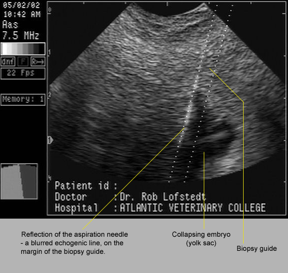
Image size:571 x 542px
In many cases, co-twins are not seen and pregnancy progresses. This usually leads to one of several outcomes. Some are shown here.
In the image below, a vesicle containing a degenerating structure believed to be an embryo, was seen on the chorion of its 60 day old co-twin. Judging from the size of this vesicle the conceptus was probably about 40 to 45 days old at the time of its death. The age of the surviving co-twin was not recorded but was probably about 65 days.

Image size: 1569 x 986px
It is possible for a dead co-twin to co-exist with a live fetus until term. Occasionally, the small, mummified fetuses will be found within the placenta of the normal foal. In other cases, the pregnancy may not go to term. This is exemplified by the case illustrated below. The mummified fetus had obviously been dead for some time yet the surviving co-twin appeared to have been normal until shortly before it was aborted. Therefore, the reason for death was not obvious in this case. Indeed, it is tempting to suggest that abortion may have been due to an unrelated problem such as EHV1 infection.

Image size:1218 x 870px Modified from original. Copyright Copyright: Dr Cyril Stephen. Charles Sturt University. NSW. Au. cyrilstephen@gmail.com
In most cases, the cause for aborted twin pregnancies appears to be related to the presence of its a co-twin. The reason for abortion is still poorly understood but may involved immunological incompatibility, endometrial sharing and other mechanisms.
Inset images A, B and C in the amalgam below show abortions that were probably related to the fact that they were twin pregnancies. These cases occurred many years ago, long before the use of ultrasound in stud practice and before the groundbreaking work of Dr O.J.Ginther, a pioneer in our understanding of twinning. Nowadays, with optimal stud management, similar images should be rare.
In the last image (D) one can see the result of a twin pregnancy that progressed to term. Occasionally normal twins are born but most twins require intensive and expensive neonatal treatment. Even so, many of these foals die or are euthanized after prolonged treatment.

Image size: 1569 x 1989px
Interestingly, it is more common for the smaller neonate to survive than its larger co-twin. In that regard, it has been suggested that the smaller co-twin is under more stress than its co-twin, accelerating fetal maturation.
It should also be noted that twin pregnancies are also potential causes of dystocia during abortion or during birth at term. In inset C, a "wryneck" fetal malformation in the larger foal complicated that delivery more than otherwise.
A cautionary note:
Often a mare is presented with premature lactation; a strong indication that abortion is imminent. If the mare has not been under one's care from the time of conception, it is just as well to warn the owner that twins may be causing the abortion. Naturally, an effort should be made to exclude placentitis. Also, the owner's expectation as to the efficiency of EHV1 vaccination should be tempered with reality. However, the possibility of twins should not be discarded even if twin heart beats cannot be discerned on transabdominal ultrasonography on repeated examinations.